Beranda
/ Anatomy Of Chest Wall - Male Anterior Thoracic Wall Chest Muscles Labeled On White Background Skin Seratus Anterior Stock Photo 200634606 - Some of the chest wall muscles can be used as helpful anatomical landmarks.
Anatomy Of Chest Wall - Male Anterior Thoracic Wall Chest Muscles Labeled On White Background Skin Seratus Anterior Stock Photo 200634606 - Some of the chest wall muscles can be used as helpful anatomical landmarks.
Insurance Gas/Electricity Loans Mortgage Attorney Lawyer Donate Conference Call Degree Credit Treatment Software Classes Recovery Trading Rehab Hosting Transfer Cord Blood Claim compensation mesothelioma mesothelioma attorney Houston car accident lawyer moreno valley can you sue a doctor for wrong diagnosis doctorate in security top online doctoral programs in business educational leadership doctoral programs online car accident doctor atlanta car accident doctor atlanta accident attorney rancho Cucamonga truck accident attorney san Antonio ONLINE BUSINESS DEGREE PROGRAMS ACCREDITED online accredited psychology degree masters degree in human resources online public administration masters degree online bitcoin merchant account bitcoin merchant services compare car insurance auto insurance troy mi seo explanation digital marketing degree floridaseo company fitness showrooms stamfordct how to work more efficiently seowordpress tips meaning of seo what is an seo what does an seo do what seo stands for best seotips google seo advice seo steps, The secure cloud-based platform for smart service delivery. Safelink is used by legal, professional and financial services to protect sensitive information, accelerate business processes and increase productivity. Use Safelink to collaborate securely with clients, colleagues and external parties. Safelink has a menu of workspace types with advanced features for dispute resolution, running deals and customised client portal creation. All data is encrypted (at rest and in transit and you retain your own encryption keys. Our titan security framework ensures your data is secure and you even have the option to choose your own data location from Channel Islands, London (UK), Dublin (EU), Australia.
Anatomy Of Chest Wall - Male Anterior Thoracic Wall Chest Muscles Labeled On White Background Skin Seratus Anterior Stock Photo 200634606 - Some of the chest wall muscles can be used as helpful anatomical landmarks.. The azygos lobe is created when a laterally displaced azygos vein makes a deep fissure in the upper part of the lung. The muscles of the thorax are also important for the vital actions of. The past several decades have seen a marked improvement in the management and reconstruction of complex chest wall de … 2 skin of the anterior chest wall syllabus p. The thorax itself can be split up into various areas that contain important structures.
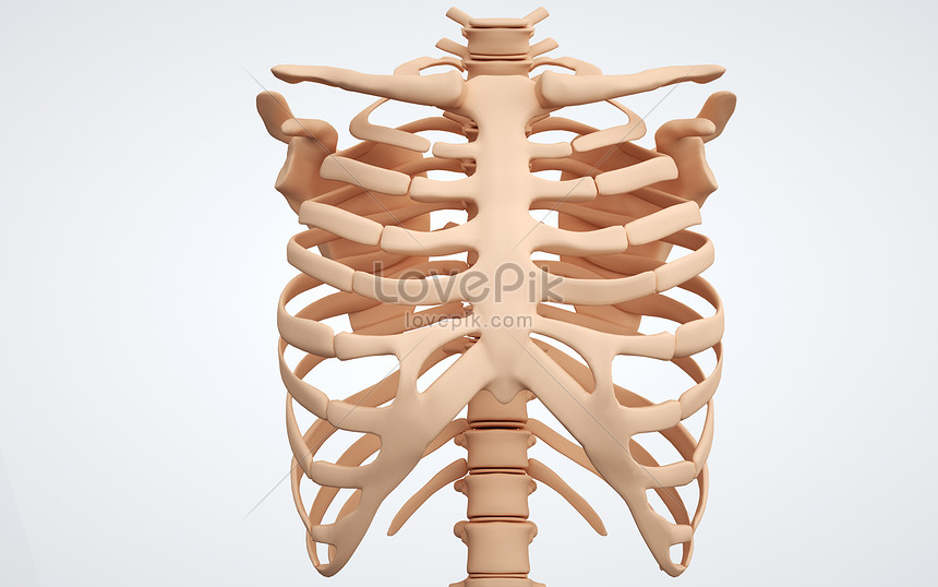
The remaining part is made up of fatty tissue. 30 lines of the thoracic wall syllabus p. The chest wall functions as a protective cage around the vital organs of the body, and significant disruption of its structure can have dire respiratory and circulatory consequences. The chest is the area of origin for many of the body's systems as it houses organs such as the heart, esophagus, trachea, lungs, and thoracic diaphragm. The chest wall is formed from the sternum anteriorly, 12 pairs of ribs, costal cartilages and intercostal muscles laterally, and the thoracic vertebrae posteriorly.
Human Chest Wall Bones Creative Image Picture Free Download 401788724 Lovepik Com from img.lovepik.com Radiological anatomy of chest including lungs,mediastinum and thoracic cage 1. Pectus excavatum is a congenital deformity of the ribs and the sternum producing a concave appearance of the anterior chest wall. 2 skin of the anterior chest wall syllabus p. The bony and soft tissue components of the chest wall combine to create an anatomic space, which houses some of the most vital structures in the human body. The azygos lobe is created when a laterally displaced azygos vein makes a deep fissure in the upper part of the lung. The thorax is bound by bony structures including the 12 pairs of ribs and thoracic vertebrae, whilst also being supported by many ligaments and muscles. We started the review by studying the thoracic wall, particularly with regard to its muscles, vessels and nerves that are of interest to mastology. The past several decades have seen a marked improvement in the management and reconstruction of complex chest wall de …
The thorax itself can be split up into various areas that contain important structures.
It provides protection to vital organs (eg, heart and major vessels, lungs, liver) and provides stability for movement. Most of the arteries of the thoracic cavity arise directly from the thoracic aorta; The chest wall, like other regional anatomy, is a remarkable fusion of form and function. The chest wall is supplied by the posterior intercostal arteries arising from the aorta, the internal thoracic and the highest intercostals given off the subclavian artery, and the branches of the axillary artery (fig. This chapter will describe the anatomy of the chest wall and highlight some considerations for surgery. The remaining part is made up of fatty tissue. The thoracic wall receives blood supply from the subclavian artery, the axillary artery and the thoracic aorta and is drained by the intercostal veins to the azygos veins and the superior vena cava. The chest wall is formed from the sternum anteriorly, 12 pairs of ribs, costal cartilages and intercostal muscles laterally, and the thoracic vertebrae posteriorly. Use the mouse scroll wheel to move the images up and down alternatively use the tiny arrows (>>) on both side of the image to move the images.>>) on both side of the image to move the images. The chest wall is comprised of skin, fat, muscles, and the thoracic skeleton. It provides access to ct images in the axial plane, allowing the user to learn and review the lung anatomy interactively. Anatomical landmarks that play an important role in clinical examination and thoracic surgery include the midsternal line, the. Chest the chest consists of bony skeleton of the spine and ribs, chest wall and diaphragm, the mediastinum and great vessels, the airways, lung parenchyma and pulmonary vessels.
The thoracic skeleton creates a protected space for the heart. The past several decades have seen a marked improvement in the management and reconstruction of complex chest wall de … It provides protection to vital organs (eg, heart and major vessels, lungs, liver) and provides stability for movement. Anatomical landmarks that play an important role in clinical examination and thoracic surgery include the midsternal line, the. It provides a protective framework for the lungs as well but also makes a significant functional contribution to respiration.
Figure 3 From Introduction To Chest Wall Reconstruction Anatomy And Physiology Of The Chest And Indications For Chest Wall Reconstruction Semantic Scholar from d3i71xaburhd42.cloudfront.net The muscles of the thorax are also important for the vital actions of. The chest wall is formed from the sternum anteriorly, 12 pairs of ribs, costal cartilages and intercostal muscles laterally, and the thoracic vertebrae posteriorly. The past several decades have seen a marked improvement in the management and reconstruction of complex chest wall de … Anatomical landmarks that play an important role in clinical examination and thoracic surgery include the midsternal line, the. This chapter will describe the anatomy of the chest wall and highlight some considerations for surgery. The thorax itself can be split up into various areas that contain important structures. The circulatory system does most of its work. The thoracic skeleton creates a protected space for the heart.
2 skin of the anterior chest wall syllabus p.
The past several decades have seen a marked improvement in the management and reconstruction of complex chest wall de … The chest wall has 10 layers, namely (from superficial to deep) skin (epidermis and dermis), superficial fascia, deep fascia and the invested extrinsic muscles (from the upper limbs), intrinsic muscles associated with the ribs (three layers of intercostal muscles), endothoracic fascia and parietal pleura. This atlas is a comprehensive and affordable learning tool for medical students and residents and especially for radiologists and pneumologists. The remaining part is made up of fatty tissue. It consists of 15 to 20 lobes of glandular tissue of the tubuloalveolar type. 6.1 diaphragm and diaphragmatic motion: How to view the anatomical labels. The circulatory system does most of its work. Chest the chest consists of bony skeleton of the spine and ribs, chest wall and diaphragm, the mediastinum and great vessels, the airways, lung parenchyma and pulmonary vessels. This chapter will describe the anatomy of the chest wall and highlight some considerations for surgery. The superior thoracic aperture found superiorly and the inferior thoracic aperture located inferiorly. It provides a protective framework for the lungs as well but also makes a significant functional contribution to respiration. The thorax has two major openings:
The azygos lobe is created when a laterally displaced azygos vein makes a deep fissure in the upper part of the lung. Arteries and veins of the thoracic wall. The thorax is bound by bony structures including the 12 pairs of ribs and thoracic vertebrae, whilst also being supported by many ligaments and muscles. The thoracic wall or chest wall is a musculoskeletal structure that has a vast vascular supply. The bony and soft tissue components of the chest wall combine to create an anatomic space, which houses some of the most vital structures in the human body.
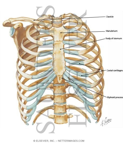
Anterior Chest Wall from www.netterimages.com It provides access to ct images in the axial plane, allowing the user to learn and review the lung anatomy interactively. Computed tomography (ct) of the chest can detect pathology that may not show up on a conventional chest radiograph(1). While others arise from its branches.on the other hand, the veins of the thoracic wall eventually coalesce to drain into the vena caval system. It furthermore supports breathing and stabilizes the shoulder girdle and upper arms during movement. The chest or thorax is the region between the neck and diaphragm that encloses organs, such as the heart, lungs, esophagus, trachea, and thoracic diaphragm. The chest is the area of origin for many of the body's systems as it houses organs such as the heart, esophagus, trachea, lungs, and thoracic diaphragm. Most of the arteries of the thoracic cavity arise directly from the thoracic aorta; The thorax is the area of the body situated between the neck and the abdomen.
While others arise from its branches.on the other hand, the veins of the thoracic wall eventually coalesce to drain into the vena caval system.
The palpable midline sternum is variable in size and shape; This mri chest (thorax) axial cross sectional anatomy tool is absolutely free to use. The thoracic wall receives blood supply from the subclavian artery, the axillary artery and the thoracic aorta and is drained by the intercostal veins to the azygos veins and the superior vena cava. The bony and soft tissue components of the chest wall combine to create an anatomic space, which houses some of the most vital structures in the human body. The thorax itself can be split up into various areas that contain important structures. Some of the chest wall muscles can be used as helpful anatomical landmarks. The thoracic wall or chest wall is a musculoskeletal structure that has a vast vascular supply. The thorax is bound by bony structures including the 12 pairs of ribs and thoracic vertebrae, whilst also being supported by many ligaments and muscles. While others arise from its branches.on the other hand, the veins of the thoracic wall eventually coalesce to drain into the vena caval system. It provides a protective framework for the lungs as well but also makes a significant functional contribution to respiration. The circulatory system does most of its work. Computed tomography (ct) of the chest can detect pathology that may not show up on a conventional chest radiograph(1). Most of the arteries of the thoracic cavity arise directly from the thoracic aorta;
Pectus excavatum is a congenital deformity of the ribs and the sternum producing a concave appearance of the anterior chest wall anatomy of chest. While others arise from its branches.on the other hand, the veins of the thoracic wall eventually coalesce to drain into the vena caval system.